Tuesday, February 24, 2009

Relapse and Leukemic Evolution: tracing back the newcomer


ResearchBlogging.org

During the last decade, the improvement of our understanding of the molecular basis of cancer has led to better diagnosis and prognosis for several types of cancer of the hematopoietic system or leukemias, as exemplified by the development of Gleevec (the commercial name of Imatinib), a drug successfully used in the treatment of acute myeloid leukemia (AML). Despite these and several other advances in cancer molecular medicine, there is no effective treatment in the event of cancer relapse and these patients have very poor prognosis. It is generally argued that relapsed leukemia is comprised of clones that display an enhanced resistance to the treatment, yet a formal comparison between the original tumors and the relapsed ones has not been performed.

In a recent issue of Science, Mullighan et al. address this problem and shed new lights on the underlying genetic basis of relapse in pediatric acute lymphoid leukemia (ALL).

By using SNP microarrays, they performed genome-wide DNA copy number analyses and compared relapsed tumors with their original counterparts (i.e. the tumor at the time of diagnosis). The samples typically showed different patterns of copy number alterations (CNAs).

By means of this genome-wide strategy, the authors were also able to validate several loci already suggested to be involved in the relapse of this disease. Besides unveiling genetic differences between the main leukemia population at the time of diagnosis and the relapsed one, they were also able to trace the origin of the latter by comparing the CNAs of the relapsed leukemia with the ones present in the leukemia at the time of diagnosis.

The logic behind this can be illustrated through an example: imagine that a relapsing leukemia lacks a chromosomal amplification that was present in the main population of cancer cells in the original tumor. This suggests that the relapsing leukemia comes from either a rare therapy-resistant clone originally present as a small subpopulation at the time of diagnosis, or it is a new leukemia altogether. If enough loci are assessed, then a “genomic-signature” (presence/absence of amplifications in determined loci) can be used as an identity tag on each leukemic clone, and from these, derive the evolutionary relationships between the different populations of tumor cells from both samples. In other words, you will be able to infer, with confidence, if the clones present in the relapse are derived from the original leukemia populations (either the main or subpopulations) or are indeed, new tumors.

Their analysis shows that 6% of relapsing leukemias are genetically distinct (i.e. possible independent tumors), 8% share the same genetic profile than the initial disease, 34% show clonal evolution from the original clone (i.e. have all the features of the initial clone, plus unique new ones), and 52% percent of the relapsed leukemias are comprised of ancestral clones that initially represented only a minor subpopulation in the original tumor.

It then appears that the majority of relapsing tumors in this disease are actually derived from a rare clone in the original tumor, further supporting the generalized notion that clinical intervention selects different subsets of cells within a heterogeneous tumor and favoring the concept of therapeutic intervention as a shaping force in the enrichment of subpopulations within a tumor. It is noteworthy that not all tumors may behave this way and it remains unclear whether these findings may be applicable to solid tumors.

Interestingly, when analyzing the genetic alterations present in the relapsed disease, no regions comprising drug resistance genes seemed to differ from their original counterparts. Provocatively, most of the alterations were present in genes involved in cell cycle regulation and B-cell development. These results challenge the view held for other leukemias, such as AML, where several studies show that relapsed diseases are comprised of drug-resistant clones.

Although there are particular known differences between “original” tumors and their relapsing counterparts, this study constitutes the first one to study genetic differences at a genome-wide level in the setting of pediatric leukemia. Their findings give new insights into the mechanisms that drive a tumor´s resistance to current therapeutic approaches and cast light onto new potential targets that could be exploited to tip the scale in favor of the relapsing patient.

The authors conclude:

"The diversity of genes that are targeted by relapse-associated CNAs, coupled with the presence of the relapse clone as a minor subpopulation at diagnosis that escapes drug-induced killing, represent formidable challenges to the development of effective therapy for relapsed ALL. Nonetheless, our study has identified several common pathways that may contain rational targets against which novel therapeutics agents can be developed".


Here's the abstract and reference:

Genomic analysis of the clonal origins of relapsed acute lymphoblastic leukemia.

Mullighan CG, Phillips LA, Su X, Ma J, Miller CB, Shurtleff SA, Downing JR.

Department of Pathology, St. Jude Children's Research Hospital, Memphis, TN 38105, USA.

Most children with acute lymphoblastic leukemia (ALL) can be cured, but the prognosis is dismal for the minority of patients who relapse after treatment. To explore the genetic basis of relapse, we performed genome-wide DNA copy number analyses on matched diagnosis and relapse samples from 61 pediatric patients with ALL. The diagnosis and relapse samples typically showed different patterns of genomic copy number abnormalities (CNAs), with the CNAs acquired at relapse preferentially affecting genes implicated in cell cycle regulation and B cell development. Most relapse samples lacked some of the CNAs present at diagnosis, which suggests that the cells responsible for relapse are ancestral to the primary leukemia cells. Backtracking studies revealed that cells corresponding to the relapse clone were often present as minor subpopulations at diagnosis. These data suggest that genomic abnormalities contributing to ALL relapse are selected for during treatment, and they point to new targets for therapeutic intervention.

C. G. Mullighan, L. A. Phillips, X. Su, J. Ma, C. B. Miller, S. A. Shurtleff, J. R. Downing (2008). Genomic Analysis of the Clonal Origins of Relapsed Acute Lymphoblastic Leukemia Science, 322 (5906), 1377-1380 DOI: 10.1126/science.1164266

Share/Save/Bookmark

Read More......

Thursday, February 19, 2009

MolBio Research Highlights joins ResearchBlogging.org


The very nature of this blog, since its origin, has been to highlight selected papers/news/tools and websites in molecular biology which we think may be interesting to the community. More recently, we have began commenting more extensively on the items we post. Yes, I can now say "we" as Francisco Barriga, a grad student at IRB Barcelona has joined this effort and will be contributing to our blog. Pancho (his nickname) is an expert on cancer biology, but as he is extremely well-read, he can make insightful commentaries on every aspect of biology. He is a great addition to the blog and I'm sure we will enjoy his participation.

In light of the path the blog has been taking recently, we decided to join a community called Research Blogging.
So, what is Research Blogging?

Do you like to read about new developments in science and other fields? Are you tired of "science by press release"? ResearchBlogging.org is your place. Research Blogging allows readers to easily find blog posts about serious peer-reviewed research, instead of just news reports and press releases.
The main idea is that scientists (or at least someone who knows what he/she is talking about) comments primary research literature on his/her blog. These serious and technical posts can then be shared through this community under a variety of categories (for example, our posts are generally posted under Biology). In general, Research Blogging readers are looking for detailed, thoughtful commentaries about research and so the submitted posts are reviewed before publication.
This site does a great service in that it helps readers find interesting and technical comments (in the form of blog posts) on articles that may be of interest to them (like many of our posts, which serve the same purpose) and also help advertise part of the myriad of science blogs out there that many of us don't know about yet.
You will be able to recognize the subset of articles we will be submitting to Research Blogging because we will place this icon on those posts:


So, take a look at the site. Who knows, you may find the science blog that you have been looking for that relates to your area of research.

Spread the word about MolBio Research Highlights, our blog.

Read More......

Wednesday, February 18, 2009

ChIP-seq accurately predicts tissue-specific activity of enhancers


by Francisco Barriga

ResearchBlogging.org
During the last decade, high throughput sequencing has given rise to an astounding amount of information regarding protein coding regions, yet very little is known about distant regulatory sequences, namely enhancers, particularly because they are scattered throughout the vast non-coding portion of the genome. In a recent issue of Nature, Visel and collaborators utilize ChIP-Seq (See [Techniques] Analyzing the genome-wide chromatin landscape: ChIP-Seq) to identify a subset of such distant-acting enhancers across the entire human genome.
[transcription][molecular biology]


They address this question by chromatin immunoprecipitation of the ubiquitously-expressed enhancer-associated protein p300 in different tissues in the mouse embryo followed by high-throughput sequencing. Interestingly the authors find that p300 binds (as a cofactor) to
different enhancer elements in the different tissues analyzed during embryo development, highlighting the importance of distant regulatory elements in appropriate gene expression and how the differential gene expression profiles may be achieved among these tissues.

Besides finding these tissue specific regions, they also validate them as bonna fide enhancers in the tissues that were predicted by p300 binding, through the use of transgenic reporter assays, showing the robustness of their findings.

An interesting fact that arose from the aforementioned studies is that p300 binding sites were generally associated with evolutionarily constrained non-coding sequences in mammalian genomes, underlying their importance in different organisms. Finally, by comparing the genomic distribution of p300 with gene expression data from the studied tissues, the authors show that there is a correlation between p300-enriched regions in embryonic tissues and the transcriptional regulation of neighboring genes, giving further functional support to their discoveries.

In conclusion, high-throughput in vivo mapping of proteins’ binding sites can then be used as an accurate means for identifying functional elements across entire genomes.

The authors conclude:

A generalized picture of the epigenetic marks and proteins associated with different types of functional non-coding elements has started to emerge from genome-wide chromatin studies. We can now begin to use these signatures to unravel gene regulation on a genomic scale in the context of living organisms.


Here's the abstract and reference:

ChIP-seq accurately predicts tissue-specific activity of enhancers.

Visel A, Blow MJ, Li Z, Zhang T, Akiyama JA, Holt A, Plajzer-Frick I, Shoukry M, Wright C, Chen F, Afzal V, Ren B, Rubin EM, Pennacchio LA.

Genomics Division, MS 84-171, Lawrence Berkeley National Laboratory, Berkeley, California 94720, USA.

A major yet unresolved quest in decoding the human genome is the identification of the regulatory sequences that control the spatial and temporal expression of genes. Distant-acting transcriptional enhancers are particularly challenging to uncover because they are scattered among the vast non-coding portion of the genome. Evolutionary sequence constraint can facilitate the discovery of enhancers, but fails to predict when and where they are active in vivo. Here we present the results of chromatin immunoprecipitation with the enhancer-associated protein p300 followed by massively parallel sequencing, and map several thousand in vivo binding sites of p300 in mouse embryonic forebrain, midbrain and limb tissue. We tested 86 of these sequences in a transgenic mouse assay, which in nearly all cases demonstrated reproducible enhancer activity in the tissues that were predicted by p300 binding. Our results indicate that in vivo mapping of p300 binding is a highly accurate means for identifying enhancers and their associated activities, and suggest that such data sets will be useful to study the role of tissue-specific enhancers in human biology and disease on a genome-wide scale.

Axel Visel, Matthew J. Blow, Zirong Li, Tao Zhang, Jennifer A. Akiyama, Amy Holt, Ingrid Plajzer-Frick, Malak Shoukry, Crystal Wright, Feng Chen, Veena Afzal, Bing Ren, Edward M. Rubin, Len A. Pennacchio (2009). ChIP-seq accurately predicts tissue-specific activity of enhancers Nature, 457 (7231), 854-858 DOI: 10.1038/nature07730

Share/Save/Bookmark

Read More......

Monday, February 16, 2009

[Techniques] Analyzing the genome-wide chromatin landscape: ChIP-Seq


By AMM

New technologies arise every few years that help us address important biological questions from a new angle. An example of this is the DNA microarray. First developed in the 1990s, this important tool has been essential for example, for profiling gene expression in specific cell types.
Nowadays, high-throughput DNA sequencing serves as an example of such important tools. In 2007, Johnson et al.1 described a technique they called “ChIP-Seq” (from CHromatin ImmunoPrecipitation and SEQuencing) to study the binding sites of a specific transcription factor across the entire human genome using the advance DNA sequencing technology from Solexa/Illumina.

Before explaining what “ChIP-Seq” is, let me tell you a little about the high-throughput technologies used before that to study the binding sites of proteins to DNA.
The most common method of locating these sites in vivo is known as chromatin immunoprecipitation (ChIP). In this technique, cells are treated with a reagent (typically formaldehyde) that crosslinks proteins to DNA. Every protein that is bound to DNA in the moment the reagent is applied will remain bound to DNA. Afterwards, the chromatin is isolated, sheared and incubated with an antibody directed to the protein of interest. This will precipitate the protein and the DNA bound by it (as they are crosslinked). After reverse-crosslinking, the precipitated DNA is analyzed. At first, this technique was restricted to studying if a particular protein was bound to a determined gene (to its promoter, protein coding region, etc), so the analysis of the precipitated DNA fragments was restricted to PCR (with the gene’s-specific primers) to see if the protein indeed allowed the precipitation of that sequence (which would result in an enrichment of the gene in the precipitated DNA vs the control, where no specific antibody is used).
About 6 years ago, a more ‘genomic’ approach was derived from this technique, resulting in the “ChIP-chip” method. The difference with the previously described technique is that after the incubation with the antibody, all the precipitated DNA fragments are used as probes on a DNA microarray (chip). This allows for a high-throughput analysis of the DNA binding sites of a specific protein (limited of course, by the sequences represented on the chip, See "Advantages" below) instead of studying single genes.

In a way to improve this technique, Johnson et al.1 replaced the “chip” by direct DNA sequencing.
What does this mean? After the precipitation and reverse-crosslinking steps, DNA fragments are sequenced (in that specific paper, using Solexa/ Illumina technology). After sequencing, the reads are mapped to the genome to determine their locations. In this way, the genome-wide DNA binding sites of a particular protein can be assessed [See figure]. The control simply omits the antibody (again, you will be looking for enrichment in your treated sample when compared to the control).
What are the advantages of ChIP-Seq vs ChIP-chip? First, you avoid all complications arising from array hybridization (probes with different optimal temperatures for binding to their complementary strands, probes that hybridize to more than one DNA sequence, interference of hybridization by DNA secondary structure). Second, you are no longer limited to what’s represented on a chip. Tiling arrays may limit such an advantage, though. Nevertheless, ChIP-Seq is cheaper (compared, for example, to whole-genome human tiling arrays)2.
Finally, you can apply ChIP-Seq regardless of whether a microarray chip has been developed for a particular species.
This technique, along with the fact that prices of high-throughput DNA sequencing are continuously becoming more accessible, will allow for the identification of the binding sites of not only transcription factors, but chromatin remodeling complexes, structural components, etc, across an entire genome.

[Image is from ref 2.]

1Johnson DS, Mortazavi A, Myers RM, Wold B (2007) Genome-wide mapping of in vivo protein-DNA interactions. Science 316, 1497.
2Fields S (2007) Molecular biology. Site-seeing by sequencing. Science. 316(5830):1441-2.


Read More......

Thursday, February 12, 2009

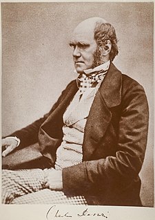
The 200th anniversary of the birth of Charles Darwin


Charles Robert Darwin was born on February 12th, 1809, in Shrewsbury, Shropshire, England.
As I have mentioned on previous posts [1][2], his contribution to both natural and social sciences has been unmatched and he is arguably one of the most important scientists of our time.
Nature will dedicate three specials to this English naturalist and has set up a website because of this special occasion, as 2009 marks his 200th birth anniversary and the 150th anniversary of the publication of the Origin of Species.
One thing I haven't mentioned is that, as would be expected, Science also set up a website (Online Collection) to commemorate this occasion, which features a blog, perspectives, research articles, etc.
Visit the site at http://www.sciencemag.org/darwin/

From the website:

The Year of Darwin

Science is celebrating the 150th anniversary of the publication of Charles Darwin's On the Origin of Species and the 200th anniversary of the author's birth with a variety of news features, scientific reviews and other special content, all collected here.


[evolution]

Read More......

Wednesday, February 11, 2009

Alternative career paths?


A few months ago, Science published an article which highlighted the bleak scenario recently (and not so recently) graduated PhDs in the life sciences face after graduation1. The size of the 'post-doc' population worldwide is dangerously increasing while the number of tenure-track academic positions is not. This generates a 'post-doc glut' which has been a problem for some time now. The number of PhDs graduating every year is by far larger that the number of positions in academia for them to fill; further, as PDFs (PostDoctoral Fellows) are 'more expensive' than grad students, many PIs tend to accept more and more grad students into their labs instead of accepting PDFs (labs need people, you know, and grants are not getting any larger). The programs themselves accept more students; where does this lead? To even more and more PhDs graduating every year and more PDFs have a hard time finding a job. Lots of offer, little demand.
Many (most) of the students enter PhD programs with the idea to follow the academic track. Are these students making informed decisions? Do they know what the job market is like? What it has to offer and what is doesn't? Has anyone taken the time to inform them about alternative career paths?
It is not, by any way, my intention to alarm any senior bachelor planning to enter a PhD program, but this is a reality. Better to know about it early.

The point is that there are alternative career paths to academia; some students even enter the programs knowing they do not want to enter that career track. Others, realize during their PhD that maybe academia is not what they want.
So, what would these alternative paths be? There are plenty of jobs in the commercial sector, science journalism, biotech industries, consulting, science administration, etc. It's not my objective to list all the possible positions, but to highlight the need to inform our students. For example, the University of Toronto has seminars by alumni who have chosen different career paths, in a way for grad students to be exposed to several options.

Should Universities accept fewer grad students? Should the efforts be made to hire more faculty? Is advice and info on alternative career paths enough? This is an ongoing debate over a very serious problem in our area of work….


See also:
Postdoc Perspective: Taking the “Alternative” out of Alternative Careers in Science
What Happened to 30 Biochemistry Graduate Students at Yale?

---

1 Mervis J. Science education. And then there was one. Science. 2008 Sep 19;321(5896):1622-8. [PubMed Link]

[ Image is from The Scientist.com ]


[career] [scientific life]

Read More......

Nature on Darwin 200


The latest issue of Nature is packed with stuff celebrating Charles Darwin's 200 birth anniversary (which falls on February 12th, by the way), but in the context of the human condition.

This is the second of three specials Nature will dedicate to this scientist with an unmatched impact on the natural and social sciences.
You can check all of the issue's content and some web-only material at the Nature's dedicated Special website www.nature.com/darwin.

From the website:
The latest edition of Nature to celebrate Darwin's life and work looks at the human side of evolution. We have features on looking for Darwin in the genome, and on what evolution has done to shape human nature, while our editorial and two commentaries look at some of the problems inherent in applying biology to questions about humanity. We also have an essay on Darwin's pigeons and poetry by his great great grand-daughter Ruth Padel. And in a special insight we bring together reviews by a range of experts on current hot topics in evolution.

Read More......

Monday, February 9, 2009

Polymerase Dynamics at the Eukaryotic DNA Replication Fork


Renowned French scientist and 1965 Nobel laureate Jacques Monod once said "What's true for E. coli is true for an elephant".
The mechanistic basis for DNA replication is partially an example of this. The extensive studies made on E. coli revealed how DNA is copied, the series of proteins involved, and how it solves the apparent 'problem' of the DNA being in an antiparallel disposition, considering that the polymerase has a polarity (the 'trombone' model. See attached video.). The mechanism (which entails recognition of the origin of replication, assembly of the replication complex, loading of the clamp, etc.) was later shown to be essentially the same in eukaryotes, although almost always involving larger complexes and more proteins (See O’ Donnell’s review for details 1).
Despite the mechanistic similarities between the DNA replication process between prokaryotes and eukaryotes, a particular difference (among many others) involves the polymerases participating in primer synthesis and elongation of the leading and lagging strand.
In E. coli, the primer is synthesized by the product of dnaG, primase, and both the leading and lagging strand are synthesized by polIII core, in the context of a holoenzyme composed of a series of subunits.
In eukaryotes, the primer on the origins (which are multiple in these organisms, in contrast to E. coli, where there is a single origin of replication) is synthesized by DNA Pol α/primase and the leading and lagging strands have been suggested to be synthesized by different polymerases; DNA polymerase{delta} would synthesize the lagging strand and DNA polymerase {epsilon} the leading.

This review discusses evidence that support this model of different polymerases acting on the leading and lagging strands and the dynamics that take place on the replication fork in eukaryotes.

Here's the abstract and references.

Polymerase Dynamics at the Eukaryotic DNA Replication Fork
Peter M. J. Burgers

Department of Biochemistry and Molecular Biophysics, Washington University School of Medicine, St. Louis, Missouri 63110

This review discusses recent insights in the roles of DNA polymerases (Pol) {delta} and {epsilon} in eukaryotic DNA replication. A growing body of evidence specifies Pol {epsilon} as the leading strand DNA polymerase and Pol {delta} as the lagging strand polymerase during undisturbed DNA replication. New evidence supporting this model comes from the use of polymerase mutants that show an asymmetric mutator phenotype for certain mispairs, allowing an unambiguous strand assignment for these enzymes. On the lagging strand, Pol {delta} corrects errors made by Pol {alpha} during Okazaki fragment initiation. During Okazaki fragment maturation, the extent of strand displacement synthesis by Pol {delta} determines whether maturation proceeds by the short or long flap processing pathway. In the more common short flap pathway, Pol {delta} coordinates with the flap endonuclease FEN1 to degrade initiator RNA, whereas in the long flap pathway, RNA removal is initiated by the Dna2 nuclease/helicase.

J. Biol. Chem., Vol. 284, Issue 7, 4041-4045, February 13, 2009


If you are interested in DNA replication, I recommend the chapter on Watson's 'Molecular Biology of the Gene' textbook. I think that chapter gives a nice intro on the concepts you need.


1 Johnson A, O'Donnell M. Cellular DNA replicases: components and dynamics at the replication fork. Annu Rev Biochem. 2005;74:283-315.

Read More......

Sunday, February 8, 2009

A novel quality-control system in protein synthesis


Protein synthesis must proceed with great accuracy, as errors may cause severe problems in the cell. Despite this need for perfection, errors are thought to occur with a rate of about 1 in 20,000 amino acids.
In the cell, the accuracy of protein synthesis is controlled by specific mechanisms at each stage.
Aminoacyl-tRNA synthetases provide the first level of control in this process, matching amino acids with specific tRNAs with an error rate of less than one per 104–105 (1).
Accuracy is further enhanced during the selection of the correct aminoacyl-tRNA by the ribosome, through its base pairing with the mRNA. This process is facilitated by a GTPase called EF1a (in bacteria, formerly known as EF-TU), or eEF1A in eukaryotes (note the extra "e", which denotes 'eukaryotes').
Kinetic proofreading (2) have been proved for both processes.

In this article published on Nature, the authors suggest the existance of a 'retrospective control mechanism' which acts after peptide bond formation (please note how the aforementioned control mechanisms act before the formation of this bond), using an in vitro bacterial translation system. A misincorporation appears to alter the translation mechanism in the ribosome somehow, increasing miscoding and also accelerating the release of the polypeptides. This is mediated by release factors, despite the absence of a stop codon.
In general, the idea seems to be that errors in translation may induce premature termination of the faulty polypeptides, in a way to avoid their synthesis. I imagine that if this mechanism indeed does take place in vivo, the early-released peptides should be rapidly targeted for degradation to avoid any chance of dominant negative effects.

As commented by Frederick and Ibba on the corresponding 'News & Views"(3)

the author's work reveals a facet of quality control in protein synthesis that depends on an unanticipated level of complexity in the workings of the ribosome

Here's the citation and abstract:

Quality control by the ribosome following peptide bond formation.

Zaher HS, Green R.

Howard Hughes Medical Institute, Department of Molecular Biology and Genetics, Johns Hopkins University School of Medicine, Baltimore, Maryland 21205, USA.

The overall fidelity of protein synthesis has been thought to rely on the combined accuracy of two basic processes: the aminoacylation of transfer RNAs with their cognate amino acid by the aminoacyl-tRNA synthetases, and the selection of cognate aminoacyl-tRNAs by the ribosome in cooperation with the GTPase elongation factor EF-Tu. These two processes, which together ensure the specific acceptance of a correctly charged cognate tRNA into the aminoacyl (A) site, operate before peptide bond formation. Here we report the identification of an additional mechanism that contributes to high fidelity protein synthesis after peptidyl transfer, using a well-defined in vitro bacterial translation system. In this retrospective quality control step, the incorporation of an amino acid from a non-cognate tRNA into the growing polypeptide chain leads to a general loss of specificity in the A site of the ribosome, and thus to a propagation of errors that results in abortive termination of protein synthesis.

Nature. 2009 Jan 8;457(7226):161-6


1Loftfield RB, Vanderjagt D. The frequency of errors in protein biosynthesis. Biochem J. 1972;128:1353–6.
2Hopfield JJ. Kinetic proofreading: a new mechanism for reducing errors in biosynthetic processes requiring high specificity. Proc Natl Acad Sci U S A. 1974;71(10):4135-9
3 Nature. 2009 Jan 8;457(7226):157-8.

Read More......

Tuesday, February 3, 2009

Cryptic Mutational Hotspots


The discovery of cryptic mutational hotspots in the human genome makes us think about the many processes underlying the determinants of mutation rates.

As discussed by Laurent Duret in the paper's1 accompanying primer2, there are neighbor-dependant mutational processes (that is, the mutation rate can vary between adjacent sites as a consequence of context). In this paper on the latest issue of PLoS Biology1, "the authors investigated the pattern of single nucleotide polymorphism (SNP) in human populations, at sites that are known to be polymorphic in chimpanzee" (see Duret's primer for a intro on the concepts discussed on the research paper), finding that there is an excess (3 times) of coincident SNPs over the number expected under the null hypothesis (which would be "SNPs are randomly distributed in the two genomes"). Interestingly, they show that this is apparently not (or at least not completely) attributable to the context and also not because of ancestral polymorphisms present in the last common ancestor of human and chimpanzee. They go on to study the possible involvement on selection to explain these conserved SNPs (positive and negative), and they conclude that the data does not support that hypothesis.

Why is this important? Briefly,

a precise knowledge of genome-wide mutation patterns is crucial for many issues in genetics (for example diseases) or evolutionary biology 2

From the authors:
We conclude that there is substantial variation in the mutation that has, until now, been hidden from view.

Here's the citation:

Cryptic Variation in the Human Mutation Rate

Alan Hodgkinson, Emmanuel Ladoukakis, Adam Eyre-Walker

Centre for the Study of Evolution, School of Life Sciences, University of Sussex, Brighton, United Kingdom

The mutation rate is known to vary between adjacent sites within the human genome as a consequence of context, the most well-studied example being the influence of CpG dinucelotides. We investigated whether there is additional variation by testing whether there is an excess of sites at which both humans and chimpanzees have a single-nucleotide polymorphism (SNP). We found a highly significant excess of such sites, and we demonstrated that this excess is not due to neighbouring nucleotide effects, ancestral polymorphism, or natural selection. We therefore infer that there is cryptic variation in the mutation rate. However, although this variation in the mutation rate is not associated with the adjacent nucleotides, we show that there are highly nonrandom patterns of nucleotides that extend ∼80 base pairs on either side of sites with coincident SNPs, suggesting that there are extensive and complex context effects. Finally, we estimate the level of variation needed to produce the excess of coincident SNPs and show that there is a similar, or higher, level of variation in the mutation rate associated with this cryptic process than there is associated with adjacent nucleotides, including the CpG effect. We conclude that there is substantial variation in the mutation that has, until now, been hidden from view.


1 Hodgkinson A, Ladoukakis E, Eyre-Walker A (2009)Cryptic Variation in the Human Mutation Rate. PLoS Biol 7(2): e27 doi:10.1371/journal.pbio.1000027

2 What is a Primer? It provides a concise introduction into an important aspect of biology highlighted by a current PLoS Biology research article.
Duret L (2009) Mutation Patterns in the Human Genome: More Variable Than Expected. PLoS Biol 7(2): e27 doi:10.1371/journal.pbio.1000027

Read More......